Fig. 1.
Download original image
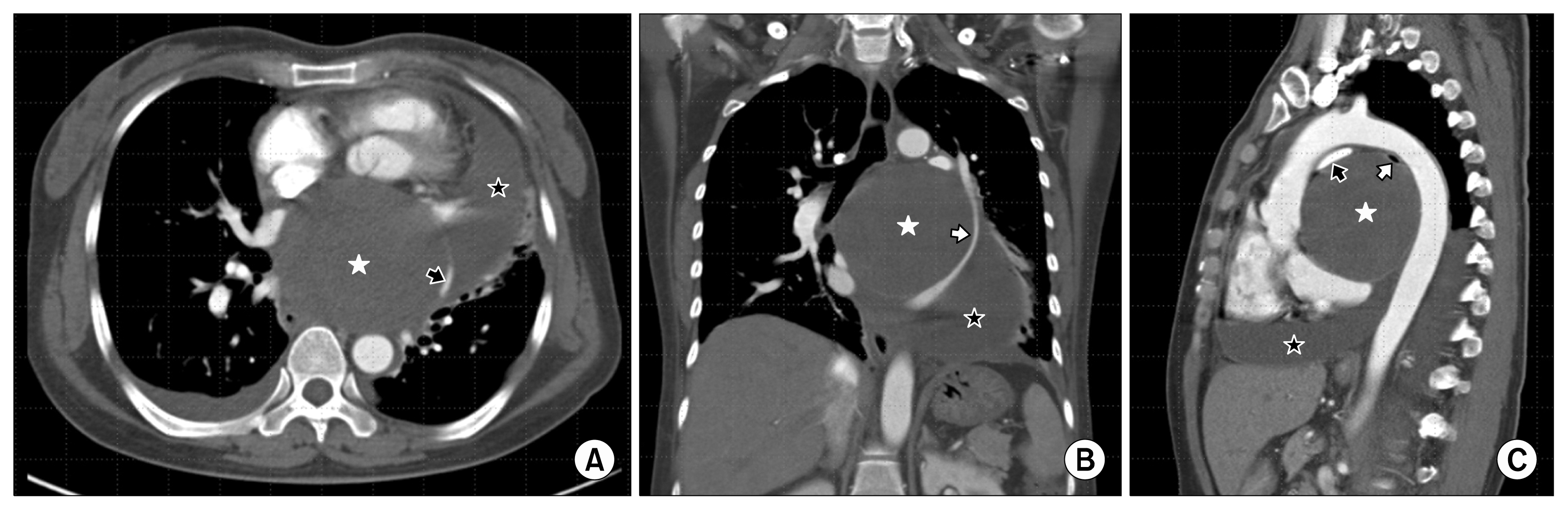
Fig. 1. (A) Computed tomography of the chest shows a very large bronchogenic cyst (white asterisk) compressing the heart and a large amount of pericardial effusion (black asterisk). The left superior pulmonary vein was compressed by a bronchogenic cyst and the distended pericardium (black arrow). (B) A coronal image shows a better view of the compressed left superior pulmonary vein (white arrow). (C) The right pulmonary artery (black arrow) and left main bronchus (white arrow) were compressed by a bronchogenic cyst and the aorta.
Korean Journal of Thoracic and Cardiovascular Surgery;51:69~71 https://doi.org/10.5090/kjtcs.2018.51.1.69
© Korean J Thorac Cardiovasc Surg